Two Channels, One Suction Path: Reading the GIF-2T1500 Clearance
Same symptom on your system?
Tell us your model and what it is doing. A GEPROBE engineer replies with a diagnosis path, part options and pricing — usually within 6 hours, with no obligation.
Chat on WhatsApp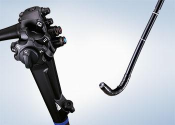
The specification sheet did not change
Olympus lists the GIF-2T1500 as an EVIS X1 dual-channel gastrointestinal videoscope, and its U.S. product page carries the same specification lines. The page for the GIF-2TH180, the dual-channel gastroscope that precedes it and the instrument Olympus compares it against, carries the same lines with the same figures.
| Specification | GIF-2T1500 (EVIS X1) | GIF-2TH180 (EVIS EXERA II) |
|---|---|---|
| Instrument channels | 2.8 mm / 3.7 mm | 2.8 / 3.7 mm |
| Working length | 1030 mm | 103 cm |
| Field of view | 140° | 140° |
| Direction of view | Forward | 0° |
| Depth of field | 2–100 mm | 2–100 mm |
| Distal end outer diameter | 12.2 mm | 12.2 mm |
| Insertion tube outer diameter | 12.6 mm | 12.6 mm |
| Angulation, up / down / right / left | 210° / 90° / 100° / 100° | 210° / 90° / 100° / 100° |
Both columns are quoted from Olympus's own pages, read on 19 October 2026. Nothing in that table is new, which is the part worth keeping: whatever the GIF-2T1500 changes, it does not change the size of the instrument, the width of either channel, the bend it can make, or the length that enters the patient. A reader who had only the sheet would be reading the specification of a scope from the previous processor generation.
Dual-channel gastroscopy is not new to the line either. Olympus describes the GIF-2TH180 as a dual-channel instrument that "can accommodate two endoscopic devices simultaneously", with the same 2.8 mm and 3.7 mm channels. So the news is not that a gastroscope has two channels. The news is in what the clearance record says about them, and that is narrower and more useful than the phrase "dual channel" suggests.
This note is written for the people who read clearance records and specification sheets as part of procurement, fleet planning or service — endoscopy unit managers, biomedical engineering, materials management. It is not clinical guidance and not a repair procedure.
What K260824 actually decided
FDA's file for the device is K260824. The clearance letter is addressed to Olympus Medical Systems Corporation and dated August 27, 2026; the submission is a traditional 510(k), Class II, under 21 CFR 876.1500, with product codes FDS and NWB. The claimed predicate is the FUJIFILM Endoscope Model EI-740D/S, K221551, and the decision recorded is "substantially equivalent".
Two things follow from that wording. The first is that this is a clearance rather than an approval: the letter states that FDA determined the device substantially equivalent to a legally marketed predicate and that "You may, therefore, market the device", which is the 510(k) route, with no premarket approval application involved. The distinction is not pedantry. An approval would rest on the manufacturer's own clinical evidence for the device; a substantial-equivalence finding rests on comparison with something already on the market.
The second follows from the first. The comparison in the file is not against Olympus's previous dual-channel scope. It is against a Fujifilm instrument, chosen by the submitter. The clearance therefore establishes that the GIF-2T1500 is as safe and as effective as the scope Olympus named — and says nothing about what changed between the GIF-2TH180 and the new model. Anyone who wants that answer has to build it from the two product pages and the device description, which is what the rest of this note does.
The summary is blunt about the evidence behind the finding: "Clinical data is not required to demonstrate substantial equivalence." What was submitted instead was bench and animal work — dimensional analysis, field of view and direction of view, depth of field, resolution, distortion, colour performance, image intensity uniformity, video latency, air, water and suction rates, thermal and photobiological safety, transport, distal attachment and detachment, plus an animal study covering narrow-band imaging, TXI, RDI and BAI-MAC observation.
Olympus and EVIS are trademarks of Olympus Corporation, and Fujifilm of Fujifilm Corporation. geprobe is an independent third-party supplier of medical equipment parts and service, with no affiliation with or endorsement from any of these companies.
Two channels, one suction path
The most useful paragraph in the file is the device description, because it assigns a job to each channel rather than counting them. In FDA's words:
The Ø 2.8 mm channel runs from the distal end to the connector and is used for inserting endoscopic accessories, introducing liquid from a syringe attached to the biopsy valve, or aspirating fluids, debris, or air from the lumen. The Ø 3.7 mm channel runs from the distal end to the instrument channel port of the control section and is used for inserting endoscopic accessories or introducing liquid from a syringe attached to the biopsy valve; this channel is not connected to the suction channel, therefore, cannot be used to aspirate the lumen.
Set that beside Olympus's own summary of the feature — dual channels "enable simultaneous use of two EndoTherapy accessories or one accessory with suction" — and the two statements agree. They agree only if you notice that they are describing different things. "One accessory with suction" is one configuration, not a property of the instrument: the accessory passes through the 3.7 mm channel and the suction comes through the 2.8 mm one. There is one suction path in this scope, and it is the smaller channel.
| Channel | Diameter | Accessories | Fluid from a syringe | Suction |
|---|---|---|---|---|
| A | 2.8 mm | yes | yes | yes — runs to the connector |
| B | 3.7 mm | yes | yes | no — not connected to the suction channel |
That table is a fair summary of the cleared description, and it changes how "accessory plus suction" should be read in a procedure plan. The configuration works when the instrument you need in the working channel is small enough for 2.8 mm and suction is the secondary task. It does not describe aspirating through the 3.7 mm channel while an accessory occupies it — a clot, a snare load, a piece of foreign body. For a case built around large-channel suction with a device in place, the record does not describe this instrument doing it.
Whether that matters is a case-mix question rather than a specification question, and it is answered by the accessories a unit actually stocks and by what its own procedures depend on. The specification table cannot answer it, and neither can the brochure sentence.
Where the change is written: the image chain
The differences FDA records between the subject device and its predicate are mostly in the imaging chain. The GIF-2T1500 uses a CMOS sensor with an RGB colour filter where the predicate uses a CCD. It offers narrow-band imaging, red dichromatic imaging and white-light imaging where the predicate offers linked colour imaging and blue laser imaging. Its depth of field starts at 2 mm rather than 3 mm, its total length is 1350 mm against 1330 mm, and its first channel is 2.8 mm against the predicate's 3.2 mm — a difference the summary addresses directly, noting that the smaller channel matches other legally marketed single-channel gastroscopes and that accessories enter and exit the image from it at an angle.
Olympus makes the same point in product terms. The scope is compatible with the EVIS X1 video system center, the CV-1500, which the FDA summary also states, and the product page lists compatibility with BAI-MAC, TXI, NBI and RDI. The imaging claim on that page is comparative: connected to the CV-1500, the GIF-2T1500 "delivers brighter images, reduced image noise, and enhanced color contrast compared to the GIF-2TH180 scope connected to the CV-190 video processor". The footnote under that sentence reads "Data on file with Olympus as of 8/27/26, 4/26/23 and 1/20/12". It is a manufacturer's comparison supported by internal data, with no study cited on the page. That does not make it untrue; it does mean a department cannot check it the way it can check a field of view.
The other caution is general. A light mode exists only on the processor generation that implements it, so a feature list for the CV-1500 is not a property of the scope by itself. This is the same distinction that decides whether two specification numbers are comparable at all, set out at more length in our note on which endoscope specifications change the device.
The reprocessing accessory is part of the clearance
The summary names a second article alongside the scope: the MAJ-420 Channel Connection Tube, "a reprocessing accessory that attaches to the GIF-2T1500 instrument channel ports and is used to inject reprocessing fluids into both instrument channels of the endoscope", and which "can also be used to flush air through both channels to expel fluids".
That is a device-specific part doing a device-specific job. Two channels are two lumens to brush, flush and dry, and the tube that reaches both at once belongs to this model's reprocessing instructions rather than to a shelf of generic fittings. A department that substitutes a connector, or loses one and improvises, is one step away from following a validated procedure on hardware the validation did not cover. The failure mode on the other end of that gap is familiar and expensive: a channel that is cleaned but not dried. What a passing leak test does and does not prove is worth reading before treating a channel check as a dryness check.
What the record does not settle
Three things a purchasing conversation tends to assume are settled by a clearance, and are not.
The first is clinical advantage. The file contains no clinical data, by FDA's own statement, and equivalence to a predicate is a regulatory finding about safety and effectiveness relative to that predicate. It is not a demonstration that procedures go better, faster or more safely with this instrument than with the one it replaces.
The second is the procedure list. Olympus's page says the dual channels are designed to support "ESD, upper GI bleeding management, foreign body removal and endoscopic suturing". The indications for use are written differently — by anatomy rather than by procedure: the instrument "is indicated for use within the upper digestive tract (including the esophagus, stomach, and duodenum)". Procedure names on a product page describe what the design is for; the labeled indication is the anatomy. Where credentialing or purchasing language needs the labeled version, that is the sentence to carry, and it is shorter than the marketing one.
The third is accessory fit. A 2.8 mm channel does not pass every accessory, and nothing in a 510(k) certifies a particular one; that check belongs to the accessory's own instructions for use.
What the record does settle is a boundary, and it is worth stating plainly. The instrument is a 12.2 mm forward-viewing gastroscope with a 2.8 mm and a 3.7 mm channel, one suction path on the smaller channel, and an imaging chain that exists on the current processor generation. If a case depends on suction through a large working channel with a device in place, this is not the instrument the record describes. That is a limit of the design rather than a defect, and telling the two apart is what the clearance document is for.
geprobe publishes no prices, lead times or stock levels, and nothing here is a quotation or a compatibility statement for any accessory. Our repair work is on endoscope-style probes — the TEE, endocavity and 4D volume transducers used with ultrasound systems — which is a different device from a gastrointestinal videoscope; nothing on this page is a statement about servicing one. What we can assess is set out on the repair page.
Related Articles
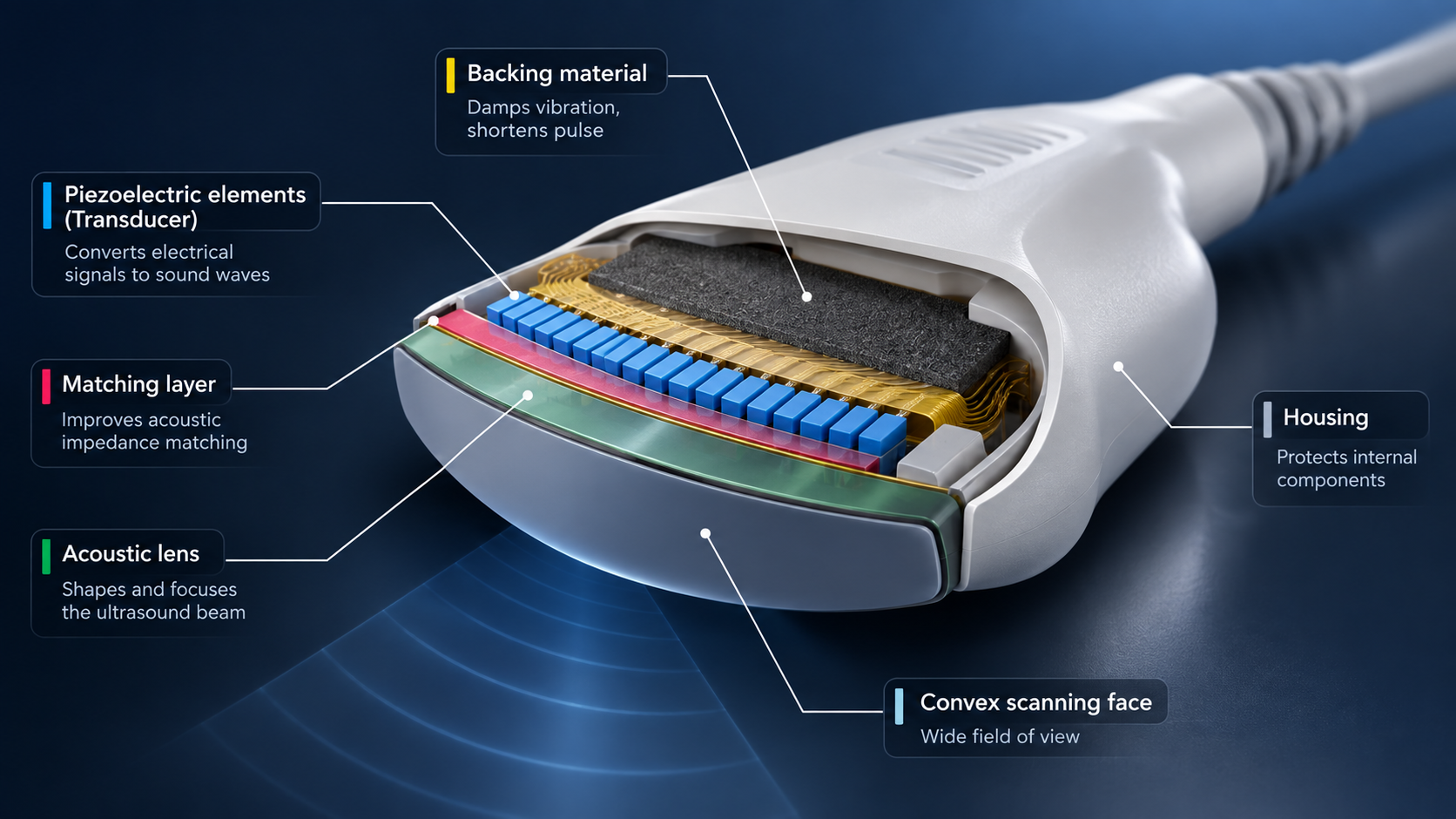
A Probe That Images Is Not Yet an Accepted Probe
A probe that images has proved one thing: it works in one port. A VIVID E9 carries two connector generations, so the receiving slip has to name the port, the six inspection points and the state at arrival.
FDA MDR Component Codes: What an Ultrasound Service Record Has to Carry
A component code names the part that was involved, not the cause. What an ultrasound service record has to carry so that someone else can code the event - and what the service side should not decide for the reporter.
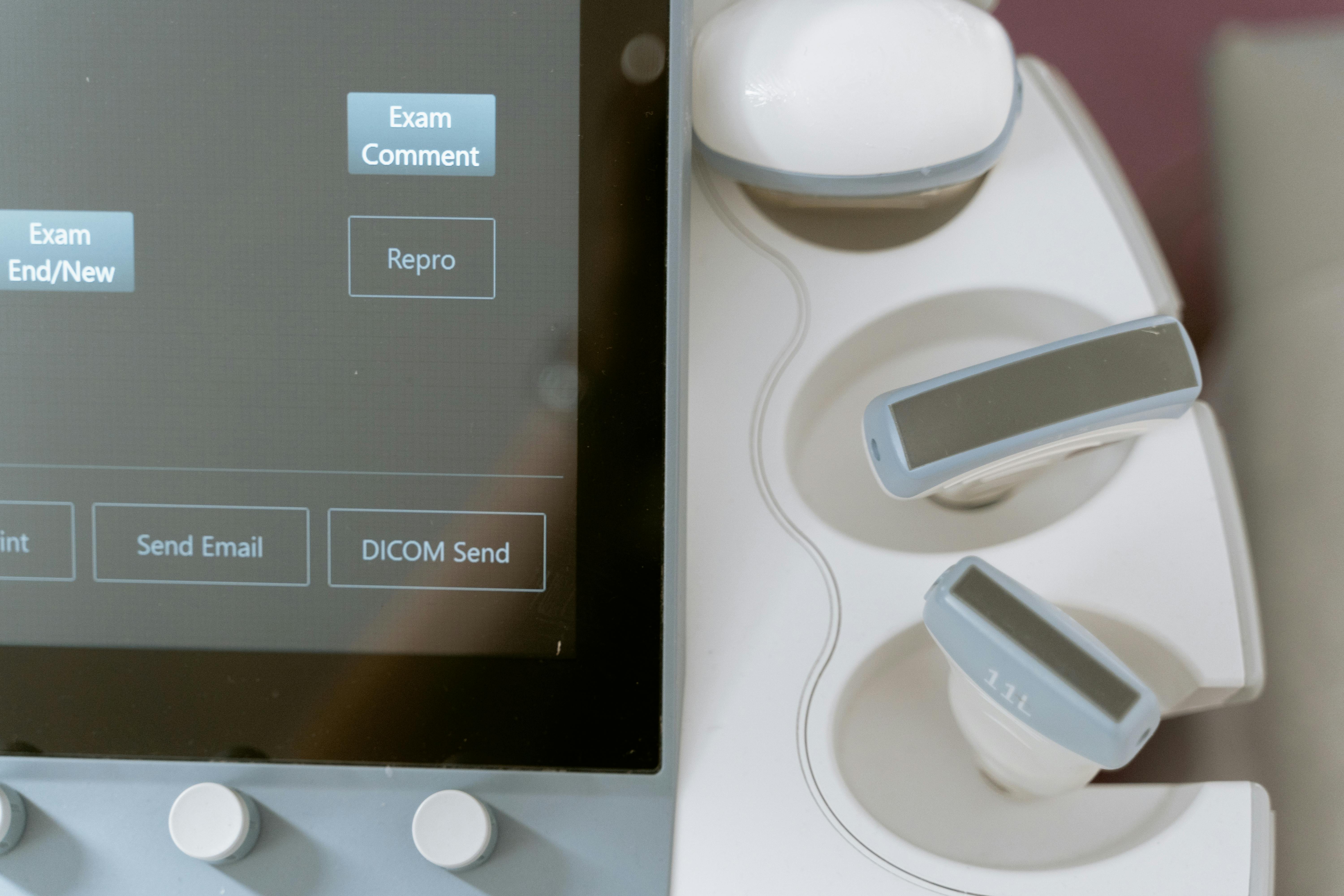
Siemens Ultrasound Needle Guides: Fitment Is Decided by the Probe
A needle guide attaches to the probe, not to the console. Which is why the deciding field on a Siemens needle guide listing names a transducer: a field-by-field read, what wears on the reusable half, and what to send before you order.